
This model is simply a representation of the HBB gene. A line is not an accurate model for the structure of genetic structures. A more accurate representation is shown below. 
0 Comments
IntroductionAs you all might remember, I wrote a post a while ago regarding the ethics of some genomic practices. Some of the questions I posed include: -Should DNA technology be used to bring back extinct species? -Is is really feasible to use DNA technology to bring back extinct species? -What are the possible health implications of DNA technology? -Could DNA technology make a truly permanent impact on the Earth as a whole? -Could DNA technology be used to eradicate disease and cancer, and if that happens, how would we sustain a planet where essentially everybody lives? In order to get a better understanding of current genome science research and hopefully answer some of the questions I posed a while back, a looked online for a current article regarding genome sciences. The article I found was this one. Summary of the Article I readThe article I read, published by National Geographic, details how scientists created human-pig chimera embryos in a lab and the implications of their work. Basically, the chimera embryos are created by taking the embryo of a pig and injecting humans stem cells into them. Early chimeras were created by taking a mouse embryo and injecting rat stem cells, some of which survived beyond the womb and actually lived on to be adults. While many protest against the hybridization of species, scientists hope to use these chimeras as donor organisms that can be used for organ transplants and the like. Scientists chose pigs as the organisms to hybridize, as their organs look a lot like human organs, even though they take less time to gestate. A challenge that the scientists will have to overcome in the future is the fact that they have to keep a low amount of human cells to pig cells. As of right now, there is about 1 human cell for every 100,000 pig cells in the chimeras. Some Questions I haveIf these human-pig embryo hybrids were able to survive beyond the lab and actually become a full grown organism, would they be subject to basic human rights?
Could the creation of a human-pig hybrid lead to another gateway for the passage of illness between species (similar to swine flu or bird flu but easier to pass on)? In our video, Vaishnavi, Olivia, Elaine, and I modeled the process of mitosis and meiosis with candy in order for our viewers to better understand the topic. While our modeling video did excelled in some aspects, such as providing an in depth explanation of mitosis, providing additional diagrams that helped deepen understanding for the viewer, and an explanation of the process of binary fission, it had some errors in the video and lacked some content that we felt we needed.
First, we never explained to our viewer that all models are wrong, and only some are useful. Our model had limitations that made it unable to fully illustrate the complexity and intricacy of mitosis and meiosis. Secondly, we never fully explained the purpose of synthesis, instead just going over the process of synthesis. We should’ve let the viewer know that synthesis is important because in order for the cell to prepare for and undergo mitosis and meiosis, all the chromosomes must be duplicated. Next, we should’ve gone more in depth into the processes and purpose of meiosis II. While we modeled meiosis II well, we didn’t explain much to the viewer and didn’t really let the viewer understand that the daughter cells divide, splitting up sister chromatids to form haploid gametes. This leads to our next shortcoming in that we didn’t really relate the process of meiosis to fertilization. We should’ve let the viewer know that the process of meiosis creates haploid gametes that fuse during fertilization to produce a diploid cell. We also should’ve modeled the process of fertilization ourselves, rather than relying on models from outside sources. Lastly, as it inevitable to have some errors in our dictation of our script, we had some slip-ups that we addressed in the description under our video. The two slip-ups that we found were that we said “centromere”, rather than centrosomes at 1:52, and we said “from meiosis”, rather than from mitosis at 5:28. Overall, I feel our group worked well together and shared the work equally. Works Cited:
"Essentials Of Genetics Unit 2: How Does DNA Move From Cell To Cell?". Nature.Com, 2014, https://www.nature.com/scitable/ebooks/essentials-of-genetics-8/126042302#bookContentViewAreaDivID. Accessed 24 Feb 2018. “Mitosis/Meiosis POGIL” IntroductionPCR, short for Polymerase Chain Reaction, is a method used to essentially copy small segments of DNA. It can be thought of as a “molecular photocopier”. Learning about PCR is important, as it is integral to DNA work because many copies of DNA are needed for molecular and genetic analyses. Materials and MethodsIn this lab, I worked with Olivia Hicks. Her blog can be found here. The first thing we did was pour the gel we would be using in the lab. We shared the gel with Olivia Studebaker, Tejal Patel, Andrew Draheim, and Nathan Washecheck. Next, we slipped on some gloves and got to the real exciting work. We put gloves on because we did not want to contaminate our reaction with our own DNA or any oils that exist on our skin. In this lab, every group that shared a gel had a control tube, which for us was prepared by Nathan and Andrew. The control tube had the same composition of the other tubes, but was not run through the thermocycler. Olivia and I prepared our tubes as follows: First, we were given our PCR tubes which we then labeled in a way that we could tell them apart. Each tube contained a little white bead, reminiscent of a Styrofoam bead, that contained the taq polymerase, nucleotides, and salt that we would need for the reaction. After labeling our tubes, we placed them in an ice bath to keep them cold. We then took them out and ensured the beads were at the bottom of the tubes by tapping them on the counter. Next, we added 20 µL of DNA primer mixture to the tubes using our trusty micropipettors and tapped them again to make sure that the primer went to the bottom of the tube. We then added 10 µL of Lambda DNA to our tube using the micropipettors and tapped our tubes once more to ensure that they had been fully mixed. The tubes were then closed and put back onto the ice, while the control was instead put next to the thermocycler. Our group then put the tubes into the thermocycler for Dr. Shingleton to run through the denature-anneal-extend cycles to prepare for the next class period. The next day, we added 4 µL of loading dye/visualization stain to our tubes so that we could be able to see our DNA under the black light. DNA doesn’t naturally glow under UV light, so the dye allowed us to see how far the DNA traveled in the lanes and analyze the results. After adding the dye, we tapped our tubes one last time to mix the contents well, and loaded each lane with about 15 µL of sample from each tube. The control, however, only used 12 µL. 15 µL of standard was also loaded into the gel. After loading, we ran the gel at 110V at 09:46. Since the loading dye had to move 45 mm to get the best results, we boosted the voltage to 120V at 10:16 and ended at around 10:20. After carefully removing our gel, (if you have read my last lab, you might remember that the gels cannot survive gravity induced trauma) we placed it in the UV light box to analyze our results. ResultsConclusionPCR is a technique used in DNA labs to essentially multiply the amount of DNA that is available for analysis. PCR is useful because it is a relatively inexpensive way to increase the amount of useful DNA needed for analysis. This lab was important to our genomics class because it taught us how to use PCR to amplify DNA and analyze it. In this lab, I learned about the process of PCR, and the importance of the thermocycler as well. As you might remember, the control was not put into the thermocycler during our experiment. As a result, we saw that the control sample only traveled 26mm as opposed to the average of 62.83 of the 6 PCR samples. This showed us that the thermocycler was integral to the process of PCR. Without the thermocycler, the reaction didn’t take place properly in the control tube. During our procedure, Olivia and I didn’t seem to make any errors on our part. However, one aspect I would like to change was the duration of the lab. It seems to me that the lab could have taken only a few hours if we were able to do it in one go. Instead, due to our school schedule, we had to complete the lab over several days, increasing the chance of contamination and error in the process. All in all, the lab was probably my favorite so far, as I felt like I was doing real DNA analysis, as opposed to the previous lab we did where we analyzed M&M dyes. One question I had while I was doing this lab was whether PCR is used in the process of cloning animals. For example, if there was only a small sample of a Wooly Mammoth’s DNA available for analysis, could PCR be used to amplify that DNA in order to preserve the original sample and allow for multiple trials? DiscussionIn the PCR technique, the DNA sample to be amplified is combined with a primer and is denatured, or separated into two pieces of single stranded DNA, through heat. This is because the heat used during PCR breaks the hydrogen bonds between the two strands of DNA in the double helix. After this, the taq polymerase enzyme builds two new strands of DNA using the original strands as a template. As a results, the new molecules of DNA that are created contain one old and one new strand of DNA. The thermocycler directs the process of denaturing and synthesis by altering the temperature every few minutes, up to 30 or 40 times to create more than 1 billion copies of the original DNA segment. Overall, PCR is a nifty technique that creates many copies of a sample of DNA, of which can be used for analysis. While DNA replication in a cell and DNA amplification by PCR are similar in the fact that they create duplicate strands of DNA, both break hydrogen bonds in the beginning of the process, and both uses primers, it is important to note the key differences between the two. DNA replication takes place in a cell at a much lower temperature than PCR. In replication, the DNA helicase enzyme “unzips” the strand of DNA to begin the process. In PCR, heat is used to break the hydrogen bonds between the strands since DNA helicase is not easily reproduced. In DNA replication, an RNA primer is used to give polymerase something to bond to. Much like in replication, a primer is used in PCR, however a DNA primer is used as opposed to an RNA primer. In replication, an RNA primer is used because the cell doesn’t have an extra DNA to use. Lastly, PCR uses taq polymerase, an enzyme found in boiling mud pits. Taq polymerase is used in PCR due to the high temperatures (up to 94°C) at which the DNA denatures and the synthesis must occur. Using polymerase from our own human body wouldn’t be feasible because the temperature would be well out of range for the functioning temperature of the enzyme. Before we ran our gels, we added a stain to our sample in order to see it. This is because DNA doesn’t glow under UV light naturally. In order to properly analyze our DNA, we need to see how far it traveled. Since the DNA we used was clear, it would have been impossible to see how far the samples traveled using natural light. We added to stain so that we could see how far the DNA traveled. Using the stain to analyze our DNA, Olivia and I estimated the size of the amplicon to be 1,400. The known size was 1,106. Our percent error of 21% was probably due to the fact that it was very difficult to see exactly where each band ended. This experiment greatly increased my knowledge of PCR and makes me exciting for the experiments to come! Bonus Works Cited“Polymerase Chain Reaction (PCR) Fact Sheet.” National Human Genome Research Institute (NHGRI), www.genome.gov/10000207/polymerase-chain-reaction-pcr-fact-sheet/.
DNA, short for deoxyribonucleic acid is an extremely complex molecule that contains all the information necessary to build and maintain an organism. It serves to pass on genetic information from parent to offspring, a characteristic known as heredity. DNA is composed of smaller subunits called nucleotides, of which there are four varieties. Nucleotides are made up of three main components, a 5-carbon sugar molecule called deoxyribose, a phosphate group, and a nitrogenous base. The nitrogenous base is responsible for the identity of the nucleotide. DNA is composed of four types of nucleotides: adenine (A), cytosine (C), guanine (G), and thymine (T). These nucleotides bond in pairs, with A and T only bonding to each other, and G and C bonding to each other. In total, there are over 246 million nucleotides in one chromosome, and over 6 billion in one cell. The picture below shows the structure of DNA in detail. In order for DNA to be passed on from parent to offspring, it must be replicated. 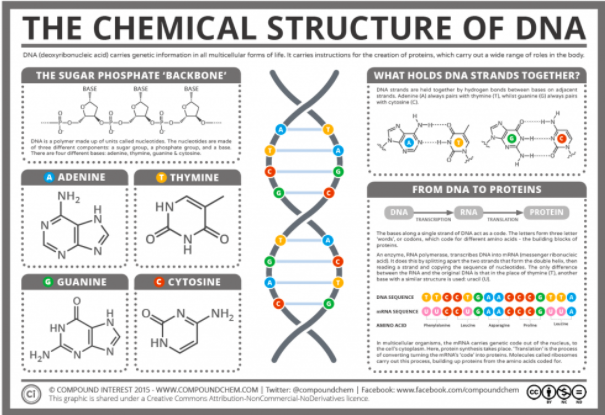  The process of DNA replication starts with helicase, an enzyme that pulls apart the double stranded DNA. It stars in an area rich with A-T pairs since these are easier to separate because they contain two hydrogen bonds as opposed to C-G pairs that contain three. Once the double stranded structure is split, two DNA polymerase enzymes collaborate to copy the leading strand and the lagging strand. On the leading strand, DNA polymerase binds the nucleotides in 5’-3’ direction, while RNA primase inserts starter RNA primer at the initial point, giving the DNA polymerase a signal to start adding the corresponding nucleotides to the strand. On the lagging strand however, RNA starts the binding process. DNA polymerase has to work backwards in 3’-5’ direction, resulting in okazaki fragments, which are short DNA segments that make up the lagging strand of the newly synthesized DNA. After finishing one okazaki fragment, the “clamp” that secures DNA polymerase to the lagging strand dissociates and lets DNA polymerase release the lagging strand temporarily. As the double stranded structure keeps getting split by the helicase, the RNA primase is initiated and inserts a short RNA primer. Then DNA polymerase clamps back to the lagging strand again and begins working from where the primers are. The polymerase stops when it reaches the point where the preceding okazaki fragment is and releases the lagging strand, waiting for RNA primase to signal. Once the copying work is done, an enzyme called exonuclease takes away the RNA primer, and DNA polymerase replaces the gap with DNA nucleotides. At the end of the process, ligase fill the gaps left in the sugar-phosphate backbone.  The structure of DNA easily facilitates the function of replication. Despite the strong double helix that stabilizes the base pairs inside the structure, the weak hydrogen bonds that hold the base pairs together allow the molecule to untwist easily for replication. The DNA molecule is split down the middle into two strands and is now able to create two copies of DNA. Various enzymes are present to stimulate the reaction as the base pairs dislodge from each other. Since the nucleotides are exposed, corresponding base pairs match up to recreate the ladder structure. Once this is complete, this newly generated DNA strand coils back up into the double helix. DNA replication is a crucial part of cell reproduction in all living organisms. In fact, life is dependent on this because without replicating DNA, our information would not pass down to future generations and life would no longer exist. Works Cited:
Introduction: In this lab, I worked with Nikhil Patel, Jackie Gibson, and Kellen Mayberry. The purpose of this lab was to get an introduction to the techniques and develop the finesse required to perform DNA labs. In preparation for this lab, Dr. Shingleton taught us how to use micropipettors. While using these instruments, I was simply amazed that it was possible for me, a clumsy teenager, to be so precise in the amount of sample I needed to use. In addition to introducing us to micropipetting, this lab taught about electrophoresis. Electrophoresis is important to DNA analysis labs because it separates macromolecules according to their size and charge. Using electrophoresis, scientists are able to analyze samples of DNA, RNA, and proteins. We used electrophoresis to analyze and identify the dyes that color M&M’s. Materials and Methods: To start off, we picked an M&M and placed it into a cup. (I picked green). We then added .5 mL of dye extraction solution to the cup and swirled it around until almost all of the dye came off of the M&M. After removing the M&M, we micropipetted the dye out of the cup and into microcentrifuge tubes. We then centrifuged our samples in order to separate the solution from the dyes. We then placed our samples into the refrigerator. We then created 4 four standards to run alongside our gel. These standards were prepared according to the table below. We then placed all of our samples into an Eppendorf rack, shown below. One thing that we unfortunately forgot to do was label our own M&M samples clearly. We decided rather the remember what color was in the tube and the position of the tube. This could have led to confusion, and is definitely something I will remember to do in the future. We also created our gel, using melted 1% agarose. We wrapped electrical tape around the gel rig and placed the comb in the middle. After this, we had Dr. Shingleton carefully pour the gel in. Once the gel was set, we tried to move the gel to the electrophoresis chamber and had an incident. We learned the hard way that gel cannot survive gravity induced trauma. The gel unfortunately fell and broke, leaving only 3 of our wells still intact. Because of this, we were not able to fully analyze our data and had to use the data of our classmates. Despite the damage done to our gel, we still loaded it in order to get the experience and develop the finesse required for our later labs. After loading the gel and setting up the rig, we put the lid on and connected the power. We ran our gels at 100 V for 15 minutes. After running our gel, we placed it on a UV light so that we could see the results. Results (Results taken from Jack Grossman, Liam Shingleton, and Olivia Hicks due to the fact that agarose gel cannot survive gravity induced trauma.) Conclusions
Using the data from the other group, I can see that Yellow 5 dye traveled the furthest, while the Blue 1 dye traveled the least. This tells me that the Yellow 5 dye is more negative and is a smaller molecule than Blue 1 dye. I can conclude this because agarose gel is like a sponge, allowing smaller particles to travel through it more easily. I know both dyes are negative because they traveled in the same direction, but Yellow 5 is more negative because it traveled further. Based on the data, it is probable that t Blue M&M contained the Blue 1 food dye. The a Green M&M contained yellow 5, and according to the group I got the data from, it might be possible it contained Blue 1 but it might be due to a spill. The Orange M&M contained either Yellow 6 and/or Red 40. It was hard to tell what it could have contained due to the lack of separation of the dyes. One thing that could have benefitted the experiment is if it were possible to create larger, more separated gels that could run for longer times. Using those larger gels, it would be easier to see the separation between dyes and also limit any cross contamination that could have happened. Discussion The gel we used in our electrophoresis is agarose. It is commonly used in the DNA research world because it is very effective at acting as a “net”, allowing small molecules to pass through it faster. This is due to the fact that when it is more like a solid, it is a porous matrix. This allows for separation between molecules with different molecular weights., measured in Daltons. For example, if there were DNA molecules with weights of 600, 1000, 2000, and 5000 Daltons, the 600 Dalton molecule would travel the farthest because it has the least molecular weight. Gel electrophoresis also separates and moves the molecules using charge. There is a positive and negative end of the gel, and depended on the charge of the molecule, it could migrate either way and any distance depending on its charge. In our lab, we only used 4 common food dyes, however, many others exist! They include the ones shown below. Sadly, we could not have used Betanin, Citrus red 2, and Carminic acid because they have no formal charge, and thus, would not move in our gel. We could use Fast green FCF because it has a negative formal charge and would move in our gel. Gel electrophoresis is a fascinating tool that scientists can use in forensics, genetics, and biochemistry. After doing this lab, I feel prepared to take on what the rest of Genomics class throws at me! Works Cited Freeman, Mary. “What Can Gel Electrophoresis Be Used For?” It Still Works | Giving Old Tech a New Life, itstillworks.com/can-gel-electrophoresis-used-5122149.html. “Gel Electrophoresis.” Science Learning Hub, 20 Nov. 2007, www.sciencelearn.org.nz/ resources/2029-gel-electrophoresis. “Introductory Gel Electrophoresis.” Carolina Biological Supply Company, 2005. Song, Guo Guo-qing, and David Douches. “Agarose Gel Electrophoresis.” 2009. January 4th, 2018. The first day of Genomics class. To be completely honest, I took Genomics only because the name and course description seemed relatively interesting. I didn't choose to take it because I had a deep interest in Genomics, but I can tell you all now that the passion I have for Genomics is definitely growing. The possibilities of DNA technology are mind boggling, but there are ethical questions that need to answered and guidelines to be set before the tech hits the real world. Personally, I think that DNA technology should be used to benefit the health of the human race and restore the planet. I think line of right and wrong really gets blurred when it comes to human procreation. For example, in the future it could be possible that we have "designer babies", where the parents essentially get to design the baby that they want. I think that is a line that should never be crossed. We should not start designing babies like we design cars and computers and the like. Some questions that I have regarding genomics and bioethics include: -Should DNA technology be used to bring back extinct species? -Is is really feasible to use DNA technology to bring back extinct species? -What are the possible health implications of DNA technology? -Could DNA technology make a truly permanent impact on the Earth as a whole? -Could DNA technology be used to eradicate disease and cancer, and if that happens, how would we sustain a planet where essentially everybody lives? |
AuthorAllan Kalapura. Holland Hall class of 2019. Archives |


 RSS Feed
Photo used under Creative Commons from Kevin M. Gill
